25 Nov 2022 | Jennifer
Why would you need a foraminotomy?
A foraminotomy is a procedure that enlarges the spinal canal exit point for nerve roots. Your nerve opening may be narrowed (foraminal stenosis). Foraminotomy takes the pressure off the nerve that is coming out of the spine, which in turn reduces pain. The foraminotomy can be performed at any level of the spine. The term foraminotomy is derived from the word- foramen means hollow passage and otomy means to open.
In foraminotomy, a surgeon removes a tissue or bone that blocks the passageway and compresses the spinal nerve root, which causes pain and inflammation. Your spinal columns protect the spinal cord from injury. Brains receive sensory information from the spinal cord. The spinal cord also conveys instructions from the brain to the body. Nerves in the spinal cord send and receives this information. Through an opening between the vertebrae known as the intervertebral foramen, they leave the spinal column.
When the opening is too small, the compressed nerve can cause pain, tingling in the arms and legs, and weakness. The symptoms of a compressed nerve vary according to its location in the spinal column. During foraminotomy, the surgeon will make a cut on your back and then surgically widen the intervertebral foramen in order to remove any blockages.
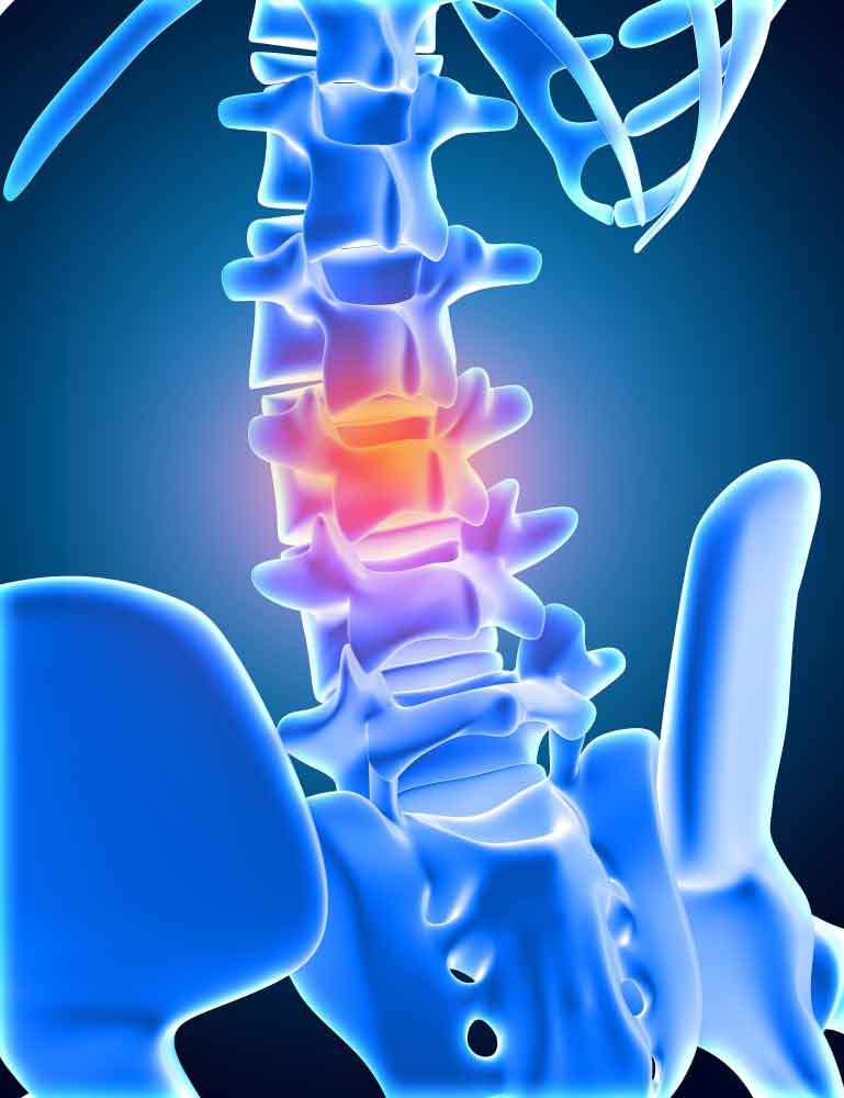
Foraminotomy is performed to release pressure from the intervertebral foramina on the spinal nerves. If you have back pain, tingling in arms or legs, or weakness, immediately contact Dallas Back Clinics.
Before the surgery
MRI and CT scan is done to make sure foraminal stenosis is the reason for your pain. Inform your healthcare provider about the medications or supplements you are taking. One week before the surgery, your healthcare provider may ask you to stop taking blood thinner medicines such as aspirin. On the day of surgery, don’t drink or eat anything for 6 to 12 hours before the procedure or after midnight the night before your procedure.
During Foraminotomy surgery
- Lay down or sit up on the operating table. Your spine is cut (incision) in the middle. The amount of your spinal column that will be operated on determines how long the incision will be.
- Ligaments, muscles, and skin are pulled to one side. A surgical microscope is used to see inside your back.
- Bone and disk fragments are moved away to open the nerve root opening.
- To provide extra space, more bone may be removed from the back of the vertebrae (laminotomy or laminectomy).
- Spinal fusion may be performed to make sure your spinal column is stable after surgery.
- The tissues, including the muscles, are repositioned. The skin is joined by sewing.

What occurs following a foraminotomy?
After the procedure :- After a couple of hours, you may be able to sit in bed. You might experience some minor pain, but medicines can help. If the surgery was on your neck, you will wear a neck collar. Follow the advice for how to take care of your back and wound. After four weeks, you should be able to resume light work again. Avoid certain movements for some time. Some people may need physical therapy as they recover. Follow all follow-up appointments. Inform your healthcare provider if no improvement is there or if you have worsening symptoms.
Should I have a foraminotomy?
Spinal stenosis blocks an intervertebral foramen or narrows the spinal column. Multiple processes have the ability to close the intervertebral foramen and compress the nerve exiting the spinal cord. Spinal stenosis can result from a number of conditions, including :-
- Spondylosis (Degenerative arthritis of the spine).
- Degeneration of the intervertebral discs.
- Enlargement of nearby ligament.
- Tumors or cysts.
- Skeletal disease.
- Congenital problems.
Nerve compression can happen in any part of the spinal column. If other treatments like physical therapy, pain medication, and injections don't work for you, you could need a foraminotomy.
What are the risks of a foraminotomy?
In most patients, foraminotomy is effective, however, occasionally problems can arise. Generally, complications of any surgery include bleeding, infection, and blood clots. Some of the possible complications are :-
- Nerve damage
- Infection in wound
- Too much blood loss
- Stroke
- Reactions to anesthesia
- Partial or no relief from pain
- The possibility of future back pain
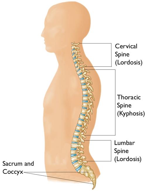
Anatomy of the spine
The cervical region (vertebrae C1-C7) consists of seven vertebrae, They provide support to the head. The atlas and axis vertebrae allow for a large range of motion in the neck, making the cervical vertebrae more mobile than other parts of the body. These vertebrae have openings that allow the spinal cord to pass through and arteries to transport blood to the brain.

The thoracic region (T1-T12) consists of 12 small bones in the upper chest. They support the ribs. Pain in this region occurs due to muscle tension, arthritis, and osteoporosis.
The vertebrae in the lumbar region (L1-L5) are much larger to support the strain of lifting and carrying heavy objects.
At the base of the spine, in the sacral area (vertebrae S1-S5) is a big bone. Five joined bones make up the triangular-shaped sacrum, which guards the pelvic organs.
If you are looking for advice or have any questions or want to book an appointment, contact us at 469-833-2927.

 Telehealth Visits Available
